
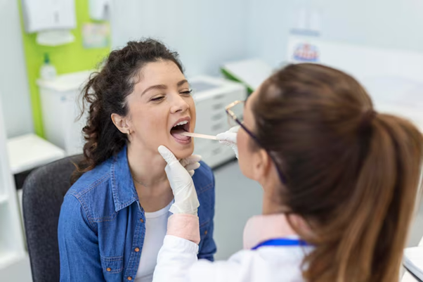
Salivary gland disorders can affect comfort, oral function, and overall health. Among these conditions, a salivary gland tumour may present with symptoms such as swelling, pain, or blockage in the salivary ducts. Early and accurate diagnosis is essential for effective management. One of the modern techniques used in evaluation is Sialendoscopy, a minimally invasive procedure that allows direct visualisation of the salivary ducts.
Understanding Salivary Gland Tumours
A salivary gland tumour can develop in any of the major salivary glands, including the parotid, submandibular, and sublingual glands. These tumours may be benign or malignant, and their symptoms can vary depending on size and location.
Common Symptoms
- Swelling near the jaw or neck
- Pain or tenderness in the affected area
- Difficulty opening the mouth
- Dry mouth or reduced saliva flow
- Recurrent infections or inflammation
Early diagnosis is important to determine the nature of the condition and guide appropriate treatment.
What Is Sialendoscopy?

Sialendoscopy is a minimally invasive diagnostic and therapeutic procedure used to examine the salivary ducts. It involves inserting a very small endoscope into the duct to provide a clear, magnified view of the internal structures.
Key Features
- Direct visualisation of salivary ducts
- Minimally invasive approach
- Can be both diagnostic and therapeutic
- Typically performed under local or general anaesthesia
This technique has become an important tool in modern salivary gland care.
How Sialendoscopy Helps Diagnose Conditions
Direct Visual Assessment
Unlike imaging tests alone, Sialendoscopy allows specialists to directly observe the inside of the salivary ducts. This helps identify:
- Blockages or narrowing
- Inflammation
- Abnormal growths or lesions
This real-time visualisation improves diagnostic accuracy.
Identifying Tumour-Related Changes
While not all tumours are located within the ducts, Sialendoscopy can reveal signs that may suggest a salivary gland tumour, such as:
- Irregular tissue growth
- Compression or obstruction of ducts
- Changes in duct structure
These findings can prompt further investigation through imaging or biopsy.
Guiding Additional Tests
Sialendoscopy often works alongside other diagnostic methods, including:
- Ultrasound
- MRI or CT scans
- Fine needle aspiration biopsy
By providing direct visual information, it helps guide where and how further tests should be performed.
Benefits Of Sialendoscopy In Diagnosis
Minimally Invasive Approach
One of the major advantages of Sialendoscopy is that it avoids the need for more invasive surgical exploration.
Accurate And Early Detection
Early identification of abnormalities supports timely diagnosis of conditions like a salivary gland tumour, improving treatment outcomes.
Reduced Patient Discomfort
Compared to traditional surgical methods, sialendoscopy typically involves:
- Less pain
- Shorter recovery time
- Minimal scarring
Combined Diagnostic And Therapeutic Use
In some cases, the procedure can also treat certain conditions, such as removing small stones or relieving blockages, while simultaneously diagnosing the issue.
When Is Sialendoscopy Recommended?
A specialist may recommend Sialendoscopy if a patient experiences:
- Recurrent salivary gland swelling
- Unexplained pain in the gland area
- Suspected duct obstruction
- Signs that may indicate a salivary gland tumour
It is particularly useful when initial imaging results are inconclusive.
Limitations And Considerations
While highly effective, sialendoscopy has certain limitations:
- It primarily examines the ducts, not deeper gland tissue
- Not all tumours are visible within the ducts
- Additional tests may still be required for confirmation
Despite these limitations, it remains a valuable part of the diagnostic process.
Role In Treatment Planning
Accurate diagnosis is essential for determining the best treatment approach. Findings from Sialendoscopy can help guide:
- Decision-making for surgery
- Monitoring of benign conditions
- Planning for further imaging or biopsy
- Minimally invasive interventions where possible
This ensures a more personalised and effective treatment plan.
Recovery And Safety
Sialendoscopy is generally considered safe, with a low risk of complications.
What To Expect
- Mild discomfort after the procedure
- Quick recovery time
- Minimal downtime
Most patients can return to normal activities shortly after the procedure.
Conclusion
Sialendoscopy has become an important tool in diagnosing salivary gland conditions. By allowing direct visualisation of the ducts, it improves the detection and assessment of abnormalities that may be associated with a salivary gland tumour.
Although it may be used alongside other diagnostic methods, its minimally invasive nature, accuracy, and ability to guide treatment decisions make it a valuable option in modern healthcare. Early evaluation using advanced techniques like sialendoscopy can lead to better outcomes, improved patient comfort, and more effective management of salivary gland conditions.